Product Code: PDBAB_016289231
The heart's rhythm is captured by a tiny, wearable device called a Holter monitor. It is utilized to identify or assess the possibility of irregular heartbeats (arrhythmias). If a conventional electrocardiogram (ECG or EKG) doesn't reveal enough information regarding the state of the heart, a Holter monitor test may be performed. Over a 24-hour period, the device tracks fluctuations in blood pressure. Some people may suffer from illnesses like heart blocks, which can cause fainting, a slowed heartbeat, and dizziness. Some people may experience atrial fibrillation or other arrhythmias that cause abnormally rapid heart rates. These problems can be identified and diagnosed using holster monitors.

Product Code: PDBAB_204019361
A test to examine your internal organs is an endoscopy. An endoscope is a long, thin tube with a tiny camera inside that is inserted into your body through a natural orifice, like your mouth. A plastic mouth guard may be required for the patient to keep their mouth open. The endoscope is then placed within the mouth. If you exhibit specific symptoms, your doctor may recommend that you get an endoscopy. Typically, a hospital's endoscopic department will perform it. Diseases that require ENDOSCOPY · Esophagus · Stomach · Upper intestine or duodenum.

Product Code: PDBAB_319055892
An electrocardiogram (ECG) is a quick test that can be used to examine the electrical activity and rhythm of your heart. The electrical signals that your heart beats out each time it beats are collected by sensors that are affixed to your skin. To check for various cardiac diseases, an electrocardiogram (ECG or EKG) measures the electrical signal from the heart. Your heart should beat evenly between 60 and 100 times per minute if the test results are normal. A fast, slow, or aberrant heart rhythm, a heart defect, coronary artery disease, heart valve disease, or an enlarged heart are just a few of the numerous cardiovascular disorders that can be detected on an ECG. EASTMED Hospital & Diagnostics PVT LTD is able to provide E.C.G. reports at affordable prices.

Product Code: PDBAB_323999793
The employees at Eastmed Hospital & Diagnostics PVT LTD are efficient and knowledgeable about the value of accurate identification, including names, dates, and times. The area of medicine known as pathology deals with the consequences of a particular disease. Samples are initially gathered and thoroughly examined while being subjected to the prescribed test. We can consistently serve all patients with precise diagnoses because of the strength of technological advancements. Pathologists focus intently on diagnosing the blood, urine, saliva, and other bodily wastes and are commonly employed in diagnostic settings. Our pathology section keeps the samples that are taken from diverse people at the proper temperature prior to the evaluation to provide accurate readings.

Product Code: PDBAB_328417441
A diagnostic procedure called electromyography (EMG) is used to evaluate the condition of the muscles and the nerve cells that govern them (motor neurons). Your herniated disc or a neurological condition like ALS or carpal tunnel syndrome may be present if your EMG reveals abnormal electrical activity when a muscle contracts. Your doctor will discuss any additional tests or treatments that might be required with you in light of your test results. The results of an EMG can identify difficulties with nerve-to-muscle signal transmission, muscle dysfunction, or even both.

Product Code: PDBAB_490583115
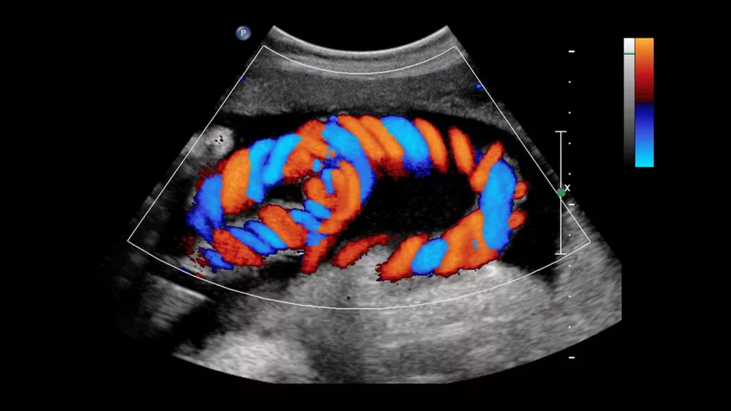
Doctors can plainly see how blood flows through the heart and blood arteries thanks to Colour Doppler ultrasound testing. Additionally, it enables them to observe and quantify artery obstructions as well as gauge the degree of heart valve leakage or constriction. Patients with atherosclerosis or coronary artery disease might be advised to take it. A Doppler ultrasonography is a painless, non-invasive method that doesn't subject you to radiation danger. This test carries no dangers, and the majority of individuals report minimal to no discomfort. Benefits · Clots of blood. · Faulty valves in your leg veins may result in blood or other fluids pooling in your legs (venous insufficiency). · Congenital heart disease and heart valve problems. · An obstructed artery (arterial occlusion).

Product Code: PDBAB_735722718
It is advised to get an NCV test done if you encounter pain or discomfort in your muscles or nerves. The following common nerve conditions can be identified using this test: An Electromyography (EMG) test and a Nerve Conduction Velocity (NCV) test are thorough and precise examinations that will assist your doctor in evaluating whether a patient has muscular or nerve damage. These are commonly carried out simultaneously. Typically, damage to these nerves results in muscle weakening, excruciating cramps, and uncontrollable muscle twitching. A patient might feel a number of symptoms since these nerves transmit information about touch, warmth, and pain. These consist of tingling or numbness in the hands or feet.

Product Code: PDBAB_738321504
A noninvasive test called an electroencephalogram (EEG) captures the electrical rhythms in your brain. Additionally, it can be utilised to verify brain death. An EEG is primarily used to identify and research epilepsy, a disorder that results in recurrent seizures. An EEG will assist your doctor in determining the type of epilepsy you have, any potential causes of your seizures, and the best treatment plan for you. Less frequently, an EEG may be used to look for other issues, such as dementia. The test is used to assist in the identification of disorders such as: · Seizures · Epilepsy · Head trauma · Vertigo · Headaches · Brain tumours · Sleeping issues.

Product Code: PDBAB_820930658
An echocardiography examines the chambers and valves of your heart to determine how effectively they are pumping blood. An echocardiogram employs ultrasound technology to view the flow of blood through the heart and electrodes to assess your heart rhythm. Your doctor can diagnose heart conditions with the aid of echocardiography. Although both ECGs and echocardiograms monitor the heart, they are two different procedures. Using electrodes, an EKG searches for aberrations in the electrical impulses of the heart. An ultrasound is used during an echocardiogram to check for structural anomalies in the heart. The echocardiography may reveal abnormalities in the heart's walls supplied by those arteries if arterial blockages are suspected.

Product Code: PDBAB_879024592
Images created by digital X-rays have greater resolution, clarity, and detail. An instant digital radiographic image is generated using digital radiography (DR), an enhanced method of x-ray inspection. With this method, data is recorded while an object is being examined using x-ray-sensitive plates, and it is then instantly transferred to a computer without the necessity of a middle cassette. Not only can better clarity aid in more accurate diagnoses and the formulation of your treatment strategy, but photographs can also be digitally edited or brightened utilizing software which results in better visibility.

Product Code: PDBAB_949747609
The uterus and ovaries can be seen throughout pregnancy, and the health of the unborn child is monitored. These are only two of the many uses for ultrasound. Like identifying the gallbladder condition or assessing the blood flow. The most frequent targets of an external ultrasound scan are the heart or an embryo in the womb. The liver, kidneys, and other organs in the abdomen and pelvis can also be examined, as well as any other organs or tissues that can be seen through the skin, like muscles and joints. Typically, the patient must fast for 8 to 12 hours prior to an abdominal ultrasound. We refer to this as fasting. Fasting assists in preventing gas buildup in the abdomen, which could influence the outcomes. Get in touch with us for more.

Product Code: PDBAB_955701055
PFT's (pulmonary function tests) are noninvasive examinations that demonstrate how effectively the lungs are functioning. The tests assess things like lung capacity, flow rates, and volume. It gauges the lungs' capacity to hold air as well as how quickly air enters and exits the lungs. Additionally, it calculates the amount of oxygen consumed and carbon dioxide exhaled while breathing. If you experience a symptom termed dyspnea, which is defined as shortness of breath, you could be advised for PFT. We draw the conclusion that the evaluation of cardiac patients for LHF may benefit from objective, accurate, and meaningful information confirmed from pulmonary function testing.